Your Blind Spot Explained: The Hole in Vision You Never Notice

How each eye loses a piece of the world-and perception quietly completes it
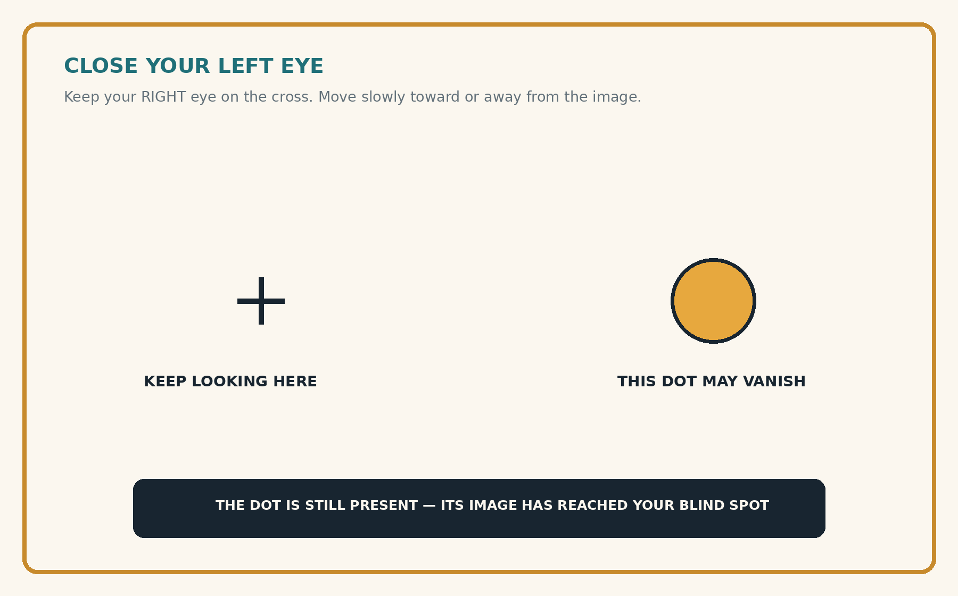
Close your left eye, fixate the cross with your right eye and move slowly toward or away from the image. At one distance, the gold dot may disappear. Original editorial demonstration.
Before You Read: Find the Missing Piece in Your Vision
Close your left eye and keep your right eye fixed on the cross. Do not look directly at the gold dot. Slowly move closer to or farther from the image. At a particular distance, the dot should disappear; continue moving, and it returns.
For the opposite eye, mirror the arrangement: close the right eye, fixate a cross on the right and place the target to its left. Screen size and viewing distance alter where the disappearance occurs, so patience is more useful than forceful staring.
WHAT JUST HAPPENED
The dot’s image fell on the optic disc-a retinal region with no photoreceptors. You did not experience a black hole. The surrounding background appeared to continue through the missing location.
What Is the Physiological Blind Spot?
Every healthy eye has a physiological blind spot: a region of the visual field corresponding to the optic disc, where retinal ganglion-cell axons gather and leave the eye as the optic nerve. Blood vessels also enter and leave at this point. Because the optic disc contains no rods or cones, light landing there cannot be detected locally.
The blind spot lies roughly 15 degrees to the temporal side of fixation and slightly below the horizontal meridian, although its exact outline varies. It is a normal anatomical feature-not a disease and not evidence that vision is defective.
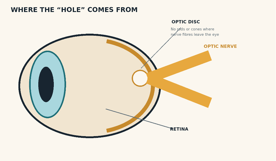
The physiological blind spot is the visual-field consequence of the optic disc: the exit point of the optic nerve contains no photoreceptors.
Original anatomical illustration.
Why Do You Never Notice It?
1. The other eye usually covers the missing region
The blind spots of the two eyes occupy different locations in visual space. With both eyes open, the fellow eye normally receives information from the area missing in the other eye. Binocular overlap therefore provides an immediate source of real visual detail.
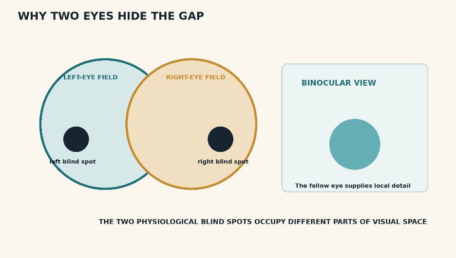
Each eye has a blind spot, but the two gaps do not coincide. Binocular overlap allows one eye to supply information missing from the other.
Editorial explanatory graphic.
2. Monocular vision still does not reveal a blank patch
Close one eye and the gap remains physically real, yet perception usually stays continuous. Colour, brightness, contours and textures surrounding the blind spot influence what is experienced there. A line crossing the region may appear uninterrupted; a uniform field may look uniformly coloured; a repeated texture may seem to continue.
3. Attention is not naturally drawn to missing input
The blind spot has no sharply visible border in ordinary viewing. Eyes, head and objects move, scenes contain redundancy, and perception is organised around surfaces and objects rather than a pixel-by-pixel inventory. Without a carefully arranged monocular test, there is little reason for the gap to announce itself.
What Does “Filling-In” Really Mean?
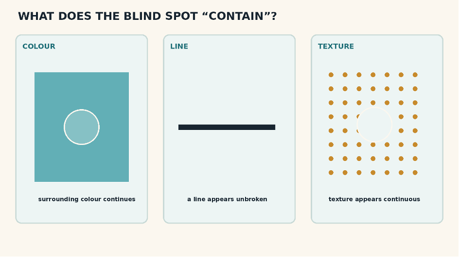
What is experienced at the blind spot depends on surrounding structure. Completion can preserve colour, contours and texture.
Editorial explanatory graphic.
“Filling-in” describes the perceptual completion of information not directly sampled at a location. The phrase should not be taken too literally. Vision may use several strategies: neural activity can extend contextual signals into the cortical representation of the blind spot, boundaries can be linked across the gap, and higher-level surface interpretations can omit the gap from conscious experience [2,5].
Experiments show that completion has rules. Simple, aligned contours and uniform surrounds fill in readily; competing patterns may produce rivalry, distortion or incomplete continuation. The visual system does not invent unlimited detail. It favours the simplest continuation supported by nearby evidence.
What Happens in the Brain?
The retina sends no direct photoreceptor signal from the optic disc, but the corresponding location is still represented within retinotopic maps of the visual cortex. Recordings in macaque primary visual cortex have identified neurons in the blind-spot representation that respond when large stimuli extend across the blind spot-conditions that support perceptual filling-in [3].
Human fMRI studies likewise show an orderly cortical map around the physiological blind spot [6]. These findings implicate early visual cortex, but they do not prove that every completed percept is produced by literal neural “paint” spreading across a map. Filling-in is better understood as a family of context-dependent completion processes [5].
A Historical Experiment Fit for a King
French physicist Edme Mariotte announced the blind spot in 1668. His demonstration used separated targets and monocular viewing so that one target vanished at the correct distance. The finding challenged the then-common assumption that the optic-nerve entrance must be the most sensitive part of the retina [1].
Historical accounts report that the demonstration was shown at the French court. By arranging a small figure’s head to fall in the observer’s blind spot while its body remained visible, the experiment could create the startling appearance of a headless person-a seventeenth-century lesson in visual anatomy.
Blind Spot, Scotoma and Troxler Fading: Not the Same Thing
The physiological blind spot is present continuously because the optic disc has no photoreceptors. A pathological scotoma is an area of impaired vision caused by disease or injury somewhere in the visual pathway. Troxler fading, by contrast, is a temporary loss of visibility for an unchanging peripheral stimulus during steady fixation; the retinal receptors are present.
All three may involve perceptual completion, but their causes differ. A normal blind-spot demonstration must never be used to dismiss a new field defect.
When a “Blind Spot” Needs Medical Attention
People often use “blind spot” loosely to describe any missing or distorted area of vision. A new, enlarging or persistent gap; a curtain or shadow; sudden visual loss; flashes with many new floaters; wavy lines; eye pain; colour desaturation; or neurological symptoms requires prompt medical or ophthalmic assessment.
Formal visual-field testing can map the normal physiological blind spot and distinguish it from abnormal defects. Persistent change is not explained simply because every eye has an optic disc.
What It Does-and Does Not-Show
It shows that the visual field is not a complete retinal photograph. Each eye contains an anatomical gap, while binocular overlap and context-dependent completion support a continuous percept.
It does not show that the brain fabricates arbitrary scenes, that peripheral vision is detailed everywhere, or that an abnormal scotoma can safely be ignored. Completion is constrained by the information that surrounds the gap.
THE TAKEAWAY
You carry a receptor-free hole in each eye, yet you almost never see it. One eye usually supplies what the other misses; when viewing with one eye, the visual system uses surrounding colour, contours and texture to preserve continuity. The blind spot is invisible precisely because perception is organised to represent a coherent world, not the gaps in its sensors.
Mini-Glossary
Optic disc - The retinal site where ganglion-cell axons form the optic nerve and blood vessels enter or leave; it contains no rods or cones.
Physiological blind spot - The normal visual-field region corresponding to the optic disc.
Photoreceptor - A rod or cone that converts light into neural signals.
Scotoma - An area of reduced or absent vision within the visual field.
Retinotopy - The orderly mapping of neighbouring retinal or visual-field locations onto neighbouring neural tissue.
Perceptual filling-in - Context-dependent completion of colour, brightness, texture, motion or contours where local visual input is absent or weak.
Evidence and Further Reading
[1] Mariotte, E. (1668). Nouvelle découverte touchant la veüe. Paris: Frédéric Léonard.
[2] Ramachandran, V. S. (1992). Filling in the blind spot. Nature, 356, 115. https://doi.org/10.1038/356115a0
[3] Komatsu, H., Kinoshita, M., & Murakami, I. (2000). Neural responses in the retinotopic representation of the blind spot in the macaque V1 to stimuli for perceptual filling-in. Journal of Neuroscience, 20(24), 9310–9319. https://doi.org/10.1523/JNEUROSCI.20-24-09310.2000
[4] Spillmann, L., Otte, T., Hamburger, K., & Magnussen, S. (2006). Perceptual filling-in from the edge of the blind spot. Vision Research, 46(25), 4252–4257. https://doi.org/10.1016/j.visres.2006.08.033
[5] Komatsu, H. (2006). The neural mechanisms of perceptual filling-in. Nature Reviews Neuroscience, 7(3), 220–231. https://doi.org/10.1038/nrn1869
[6] Awater, H., Kerlin, J. R., Evans, K. K., & Tong, F. (2005). Cortical representation of space around the blind spot. Journal of Neurophysiology, 94(5), 3314–3324. https://doi.org/10.1152/jn.01330.2004
Educational note: This article explains the normal physiological blind spot. It is not a substitute for visual-field testing or assessment of new visual symptoms.


Comments